Criteria used to evaluate the consistency of the bony and facial outlines/morphological curves
This group of criteria is based on evaluating the most common relationships between curves and / or bone contours (B) and curves and / or facial contours (F). We can distinguish three main types of relationships: F follows B; F is consistent with B; and F is the mirror image of B. For example, the contour of the frontal bone follows the contour of the forehead on the face or the contour of the chin is consistent with the mental contour.
-
Criteria assessed in Frontal View:
- The width of the cranium fills the forehead area of the face.
- The length of the skull from menton to bregma fits within the face.
- The lateral line of the zygomatic bone matches the outline of the cheek.
- The outline of the face and the outline of the skull all along the contour follow each other maintaining symmetrical flow by side.
- The outline of the nose in the face represented by shade distribution follows the outline of the nasal bone in the skull maintaining symmetrical flow by side.
- The arcus supraciliaris follows supraorbital margin.
- Oblique line of the mandible.
- Dental information (bony to bony consistency).
-
Criteria assessed in Lateral/Oblique View:
- The outline of the frontal bone follows the forehead outline.
- The skull and head height is similar.
- The chin outline is consistent with the mental outline.
- The gonial outline follows the outline of jaw angle.
- The outline of the frontal process of the zygomatic bone can be aligned with the process seen in the face.
- The arcus supraciliaris follows supraorbital margin.
- The outline of the nasal bones follows the outline of the nose in the skull with minimal tissue thickness allowance.
Criteria assessed in Frontal View
This group includes a series of anatomical criteria that can be evaluated in a frontal view photograph.
- The width of the cranium fills forehead area of the face.
 Figure 1. Example of a positive match in which the cranial width is evaluated with Skeleton·ID by means of the transparency and wipe tools showing consistency with the facial width. The width of the skull fits within the facial contour.
 Figure 2. Example of a negative match in which the cranial width is evaluated with Skeleton·ID by means of the transparency and wipe tools showing inconsistency with the facial width. The width of the skull is greater than the width of the face, protruding from the facial contour.
- The length of the skull from menton to bregma fits within the face.
 Figure 3. Example of a positive match in which the length of the skull is evaluated with Skeleton·ID by means of the transparency tool, showing that the length of the skull from menton to bregma fits within the face contour consistently.
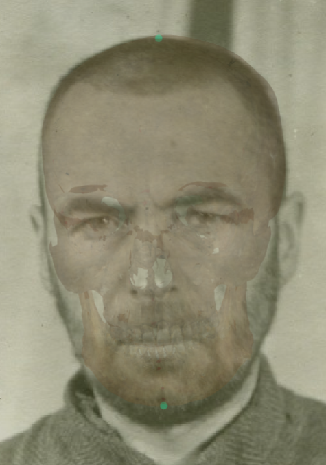 Figure 4. Example of a negative match in which the length of the skull is evaluated with Skeleton·ID by means of the transparency tool, showing that the length of the skull from menton to bregma does not fit within the face contour. The length of the skull is greater than the length of the face. Bregma is located outside the facial contour.
- The lateral line of the zygomatic bone matches the outline of cheek.
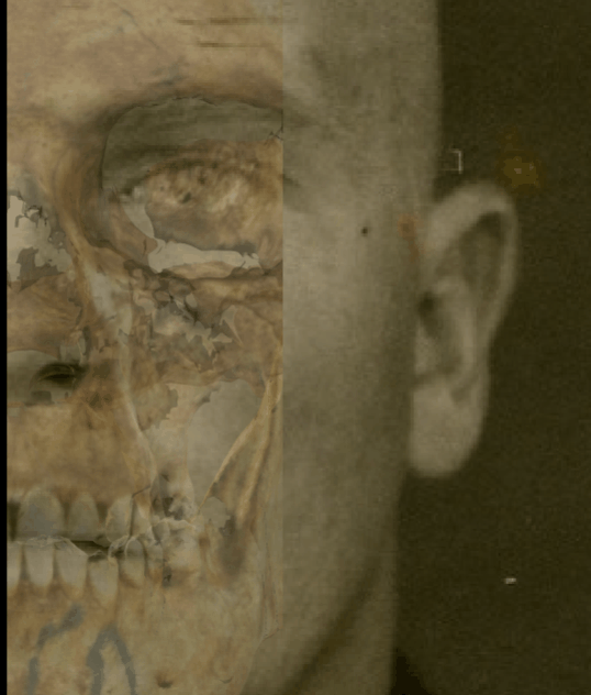 Figure 5. Example of a positive match in which the lateral line of the zygomatic bone is evaluated with Skeleton·ID by means of the transparency and wipe tools, showing that the lateral line of the zygomatic bone matches the lateral line of the cheek in the face.
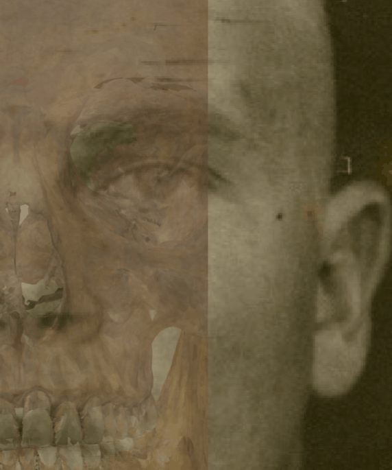 Figure 6. Example of a negative match in which the lateral line of the zygomatic bone is evaluated with Skeleton·ID by means of the transparency and wipe tools, showing that the lateral line of the zygomatic bone does not match the lateral line of the cheek on the face. The zygomatic bone does not reach the contour of the cheek on the face.
- The outline of the face and the outline of the skull all along the contour follow each other maintaining symmetrical flow by side.
 Figure 7. Example of a positive match in which the outline of the skull and face is evaluated with Skeleton·ID by means of the transparency and wipe tools, showing that the outline of the face follows the outline of the skull all along the contour maintaining symmetrical flow by side in a consistent way. The wipe tool has been used to show a gradient from right to left of the skull over the face.
 Figure 8. Example of a negative match in which the outline of the skull and face is evaluated with Skeleton·ID by means of the transparency and wipe tools, showing that the outline of the skull does not follow the outline of the face in a consistent way. The frontal outline of the skull protrudes from the facial outline. The wipe tool has been used to show a gradient from right to left of the skull over the face.
- The outline of the nose in the face represented by shade distribution follows the outline of the nasal bone in the skull maintaining symmetrical flow by side.
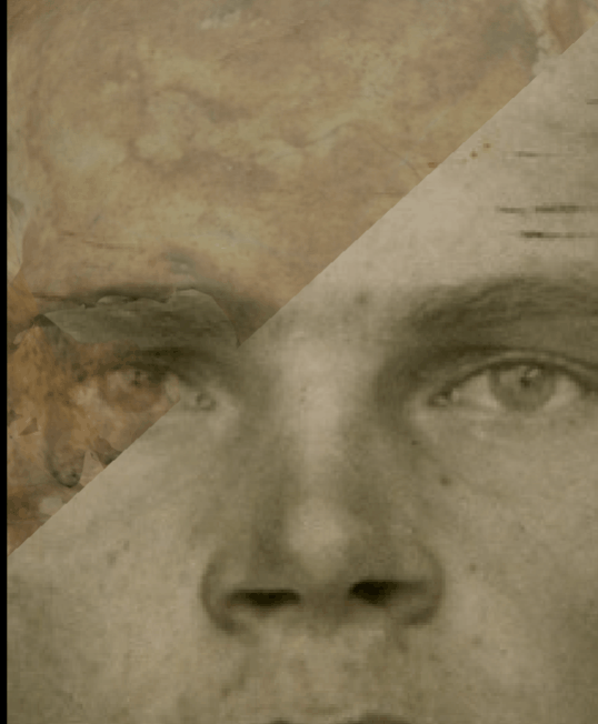 Figure 9. Example of a positive match in which the outline of the nose is evaluated with Skeleton·ID by means of the transparency and wipe tools, showing that the outline of the nasal bone maintains a symmetrical flow on both sides with the outline of the nose in the photograph in a consistent way. The wipe tool has been used to show a gradient from top to bottom and from left to right of the nasal bone over the nose.
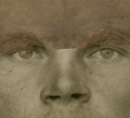 Figure 10. Example of a negative match in which the outline of the nose is evaluated with Skeleton·ID by means of the transparency and wipe tools, showing that the outline of the nasal bone does not maintain a symmetrical flow on both sides with the outline of the nose in the photograph in a consistent way. The nasal bone is larger and does not fit with the nose in the photograph. The wipe tool has been used to show a gradient from top to bottom and from left to right of the nasal bone over the nose.
- The arcus supraciliaris follows supraorbital margin.
 Figure 11. Example of a positive match in which the arcus supraciliaris is evaluated with Skeleton·ID by means of the transparency and wipe tools, showing that the arcus supraciliaris of the face follows the supraorbital margin of the skull in a consistent way. The wipe tool has been used to show a gradient from right to left (anatomical) of the supraorbital margin over the arcus supraciliaris.
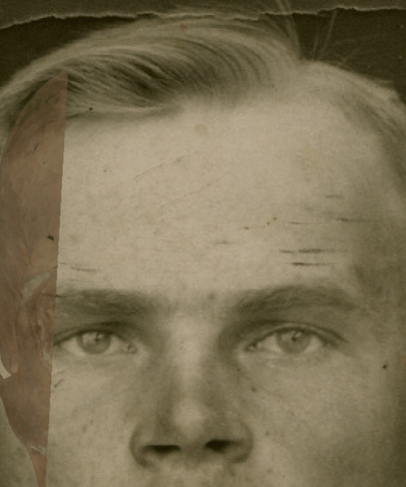 Figure 12. Example of a negative match in which the arcus supraciliaris is evaluated with Skeleton·ID by means of the transparency and wipe tools, showing that the arcus supraciliaris of the face does not follow the supraorbital margin of the skull in a consistent way. The wipe tool has been used to show a gradient from right to left (anatomical) of the supraorbital margin over the arcus supraciliaris. In this case the supraorbital margin on the skull is located above the superciliary arch on the face, showing inconsistency between both anatomical structures.
- Oblique line of the mandible. This criterion should be considered with caution due to the low discriminative power and subjectivity in frontal view according to Ibáñez y col. (2016).
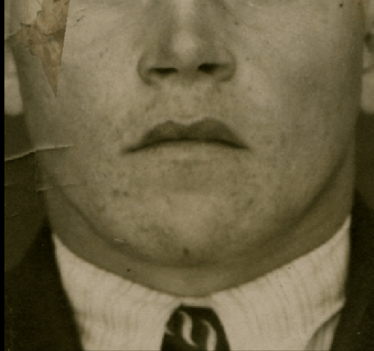 Figure 13. Example of a positive match in which the oblique line of the mandible is evaluated with Skeleton·ID by means of the transparency and wipe tools, showing that the oblique line of the mandible (left and right) on the face follows the oblique line of the mandible on the skull in a consistent way. The wipe tool has been used to show a gradient from right to left (anatomical) of the mandible on the skull over the face.
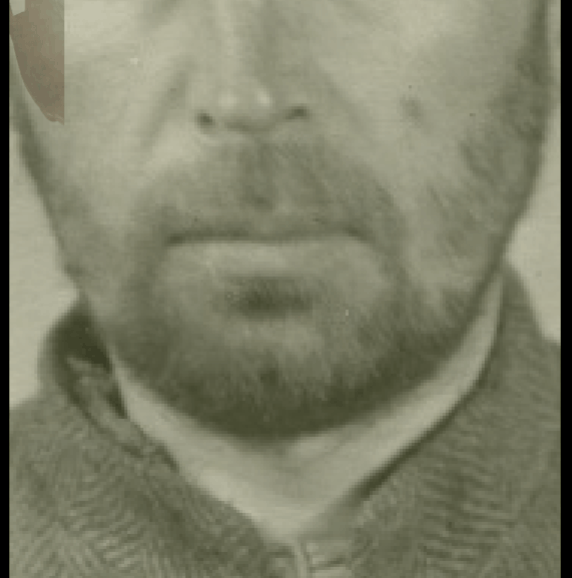 Figure 14. Example of a negative match in which the oblique line of the mandible is evaluated with Skeleton·ID by means of the transparency and wipe tools, showing that the oblique line of the mandible (left and right) on the face does not follows the oblique line of the mandible on the skull in a consistent way. The wipe tool has been used to show a gradient from right to left (anatomical) of the mandible on the skull over the face. In this case, the gonial outline is not consistent with the mandible outline of the face.
- Dental information (bony to bony consistency). This criterion has a highest discriminative power according to Ibáñez y col. (2016).
 Figure 15. Example of a positive match in which the dental consistency is evaluated with Skeleton·ID by means of the transparency tool, showing that the bony to bony consistency of the upper teeth of the skull match with the upper teeth of the photograph. The opacity tool has been used to show a gradient of transparency of the skull teeth over the facial photograph.
 Figure 16. Example of a negative match in which the dental consistency is evaluated with Skeleton·ID by means of the transparency tool, showing that the bony to bony consistency of the upper teeth of the skull does not match with the upper teeth of the photograph. The opacity tool has been used to show a gradient of transparency of the skull teeth over the facial photograph.
Criteria assessed in Lateral/Oblique View
This group includes a series of anatomical criteria that can be evaluated in a lateral and / or oblique view photograph.
- The outline of the frontal bone follows the forehead outline. This is the best criteria for the evaluation in lateral view according to Ibáñez y col. (2016).
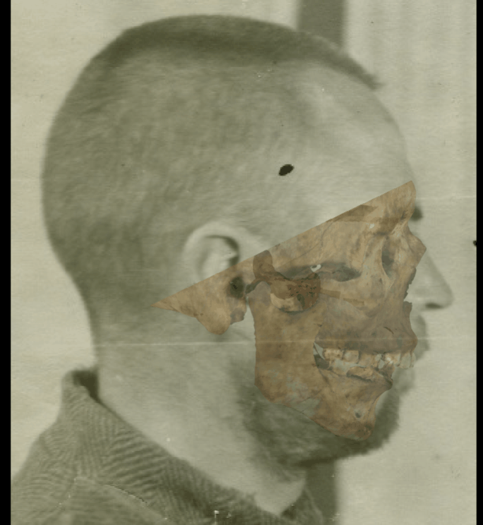 Figure 17. Example of a positive match in which the forehead outline is evaluated with Skeleton·ID by means of the transparency and wipe tools, showing that the outline of the frontal bone follows the forehead outline in a consistent way. The wipe tool has been used to show a gradient from the supraorbital margin to the forehead.
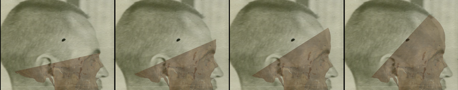 Figure 18. Example of a negative match in which the forehead outline is evaluated with Skeleton·ID by means of the transparency and wipe tools, showing that the outline of the frontal bone does not follow the forehead outline in a consistent way. The wipe tool has been used to show a gradient from the supraorbital margin to the forehead. In this case it can be seen how the contour of the forehead in the skull protrudes from the contour of the forehead on the face.
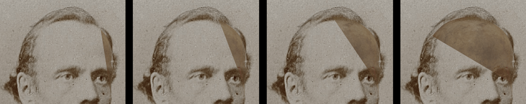 Figure 19. Example of a positive match in which the forehead outline is evaluated with Skeleton·ID by means of the transparency and wipe tools, showing that the outline of the frontal bone follows the forehead outline in a consistent way. The wipe tool has been used to show a gradient from the supraorbital margin to the forehead.
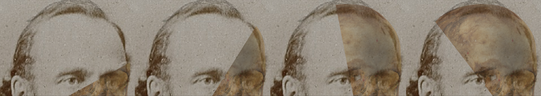 Figure 20. Example of a negative match in which the forehead outline is evaluated with Skeleton·ID by means of the transparency and wipe tools, showing that the outline of the frontal bone does not follow the forehead outline in a consistent way. The wipe tool has been used to show a gradient from the supraorbital margin to the forehead. In this case it can be seen how the contour of the forehead in the skull protrudes from the contour of the forehead on the face.
- The skull and head height is similar. The outline of the face and the outline of the skull all along the contour follow each other. This criterion has an important discriminative power and a significant variability due to the variation in soft tissue and distortion in the perception created by presence of hair.
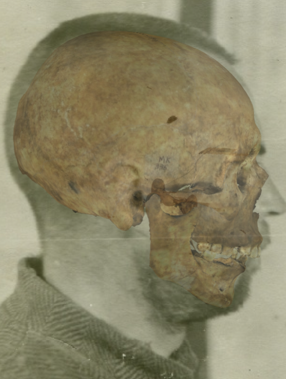 Figure 21. Example of a positive match in which the skull height is evaluated with the transparency tool, showing that the skull and head height is similar in a consistent way. In this case the distortion in the perception created by presence of hair is minimal facilitating the evaluation of this criterion.
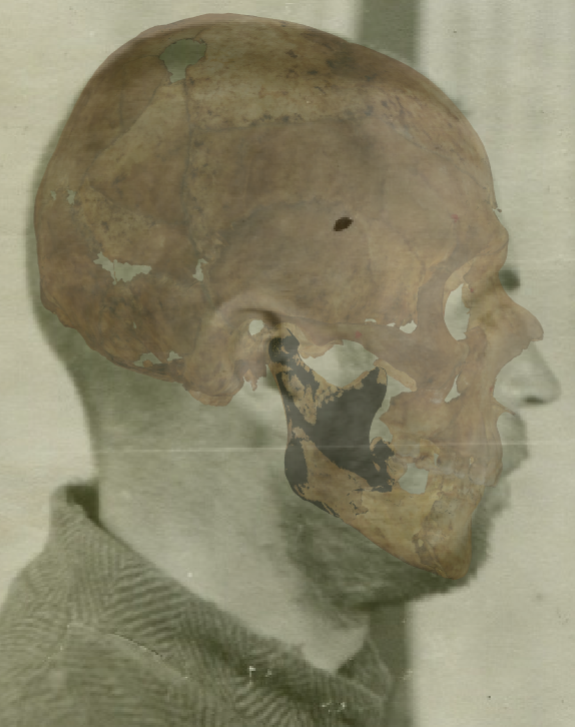 Figure 22. Example of a negative match in which the skull height is evaluated with the transparency tool, showing that the skull and head height is not similar in a consistent way. In this case the distortion in the perception created by presence of hair is minimal facilitating the evaluation of this criterion and it can be seen how the upper cranial contour protrudes from the upper facial contour so that the height of the skull is greater than the height of the face.
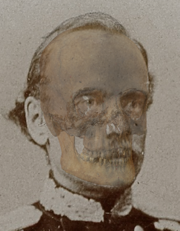 Figure 23. Example of a positive match in which the skull height is evaluated with the transparency tool, showing that the skull and head height is similar in a consistent way. In this case the distortion in the perception created by presence of hair is minimal facilitating the evaluation of this criterion.
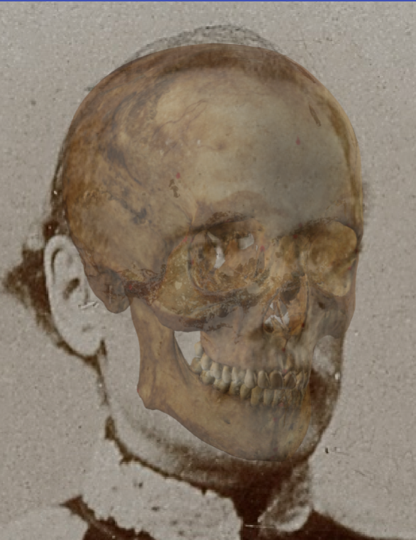 Figure 24. Example of a negative match in which the skull height is evaluated with the transparency tool, showing that the skull and head height is not similar in a consistent way. In this case the distortion in the perception created by presence of hair is minimal facilitating the evaluation of this criterion and it can be seen how the upper cranial contour protrudes from the upper facial contour so that the height of the skull is greater than the height of the face.
- The chin outline is consistent with the mental outline.
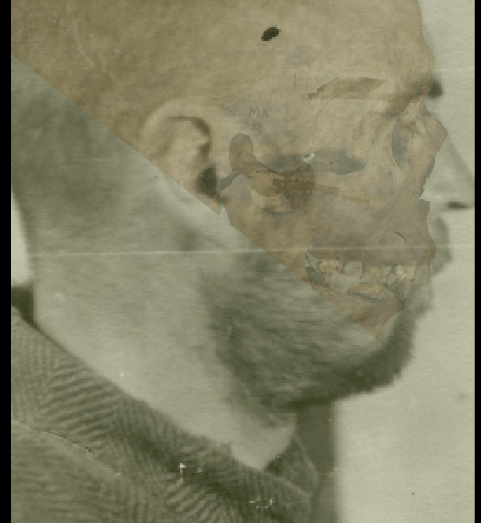 Figure 25. Example of a positive match in which the chin outline is evaluated with the transparency and wipe tools, showing that the chin outline is consistent with the mental outline. The wipe tool has been used to show a gradient from right to left (anatomical) of the mental outline (skull) over the chin outline.
 Figure 26. Example of a negative match in which the chin outline is evaluated with the transparency and wipe tools, showing that the chin outline is not consistent with the mental outline. The wipe tool has been used to show a gradient from right to left (anatomical) of the mental outline (skull) over the chin outline. In this case the mental outline protrudes from the chin outline on the face, showing an incompatible inconsistency.
 Figure 27. Example of a positive match in which the chin outline is evaluated with the transparency and wipe tools, showing that the chin outline is consistent with the mental outline. The wipe tool has been used to show a gradient from right to left (anatomical) of the mental outline (skull) over the chin outline.
 Figure 28. Example of a negative match in which the chin outline is evaluated with the transparency and wipe tools, showing that the chin outline is not consistent with the mental outline. The wipe tool has been used to show a gradient from right to left (anatomical) of the mental outline (skull) over the chin outline. In this case the mental outline does not follow until the end of the chin, turning out to be narrower showing an incompatible inconsistency.
- The gonial outline follows the outline of jaw angle. This criterion is easily to evaluate and has an important discriminative power according to Ibáñez y col. (2016).
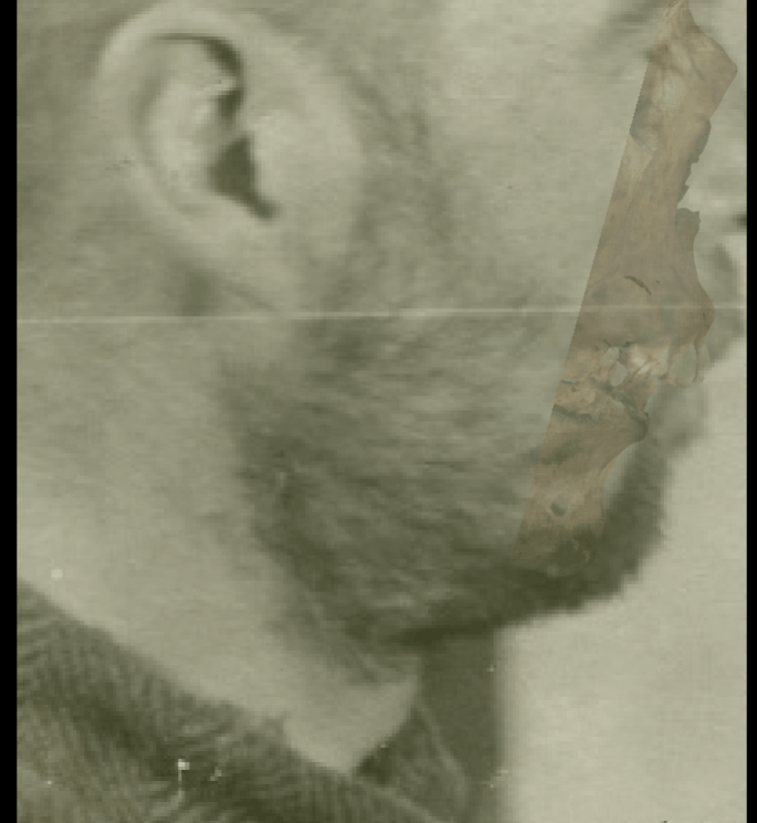 Figure 29. Example of a positive match in which the gonial outline is evaluated with the transparency and wipe tools, showing that the gonial outline follows the outline of the jaw in a consistent way. The wipe tool has been used to show a gradient from the chin to the jaw angle showing a concordance between the two anatomical structures.
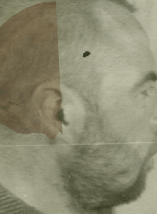 Figure 30. Example of a negative match in which the gonial outline is evaluated with the transparency and wipe tools, showing that the gonial outline does not follow the outline of the jaw in a consistent way. The wipe tool has been used to show a gradient from the mastoid process to the chin showing that the chin protrudes from the facial contour and the jaw angle is not consistent.
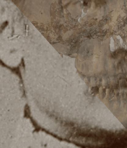 Figure 31. Example of a positive match in which the gonial outline is evaluated with the transparency and wipe tools, showing that the gonial outline follows the outline of the jaw in a consistent way. The wipe tool has been used to show a gradient from the chin to the jaw angle showing a concordance between the two anatomical structures.
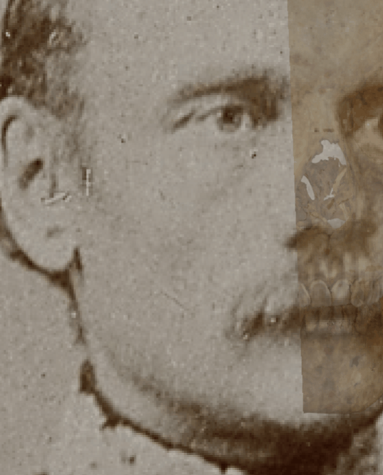 Figure 32. Example of a negative match in which the gonial outline is evaluated with the transparency and wipe tools, showing that the gonial outline does not follow the outline of the jaw in a consistent way. The wipe tool has been used to show a gradient from the chin to the jaw angle.
- The outline of frontal process of the zygomatic bone can be aligned with the process seen in the face. The outline of the zygomatic arch can be fitted between the skull and the face (these criteria are more easily appreciated in individuals with minimal soft tissue thickness).
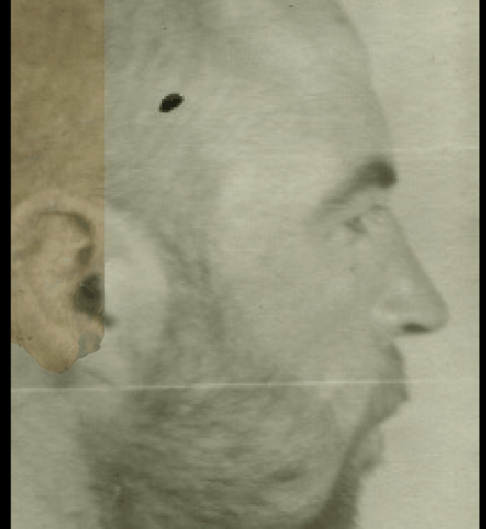 Figure 33. Example of a positive match in which the frontal process of the zygomatic arch and the zygomatic arch are evaluated with the transparency and wipe tools, showing that the frontal process can be aligned with the process in the face and the outline of the zygomatic arch can be fitted between the skull and the face in a consistent way. The wipe tool has been used to show a gradient from the mastoid process to the frontal process of the zygomatic bone.
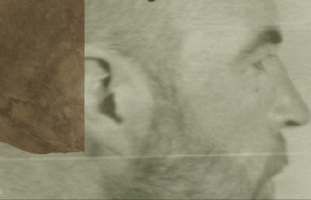 Figure 34. Example of a negative match in which the frontal process of the zygomatic arch and the zygomatic arch are evaluated with the transparency and wipe tools, showing that the frontal process is not aligned with the process in the face and the outline of the zygomatic arch can’t be fitted between the skull and the face (cause the zygomatic arch is larger than de facial width). The wipe tool has been used to show a gradient from the mastoid process to the frontal process of the zygomatic bone.
- The arcus supraciliaris follows supraorbital margin. This criterion should be considered with caution due to the low discriminative power and subjectivity in lateral / oblique view according to Ibáñez y col. (2016).
 Figure 35. Example of a positive match in which the arcus supraciliaris is evaluated with the transparency and wipe tools, showing that the arcus supraciliaris follows the supraorbital margin in a consistent way. The wipe tool has been used to show a gradient from the zygomatic arch to the supraorbital margin showing how the contour of the arcus supraciliaris follows the supraorbital margin until the end.
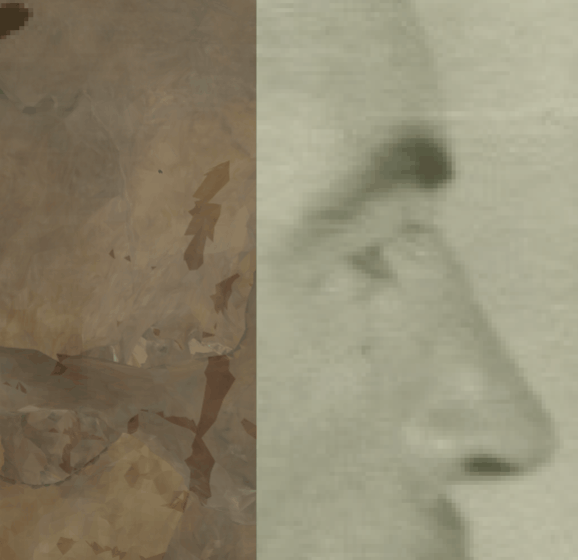 Figure 36. Example of a negative match in which the arcus supraciliaris is evaluated with the transparency and wipe tools, showing that the arcus supraciliaris does not follow the supraorbital margin in a consistent way. The wipe tool has been used to show a gradient from the zygomatic arch to the supraorbital margin showing how the contour of the supraorbital margin protrudes over the arcus superciliaris.
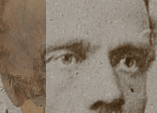 Figure 37. Example of a positive match in which the arcus supraciliaris is evaluated with the transparency and wipe tools, showing that the arcus supraciliaris follows the supraorbital margin in a consistent way. The wipe tool has been used to show a gradient from the zygomatic arch to the supraorbital margin showing how the contour of the arcus supraciliaris follows the supraorbital margin until the end.
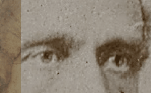 Figure 38. Example of a negative match in which the arcus supraciliaris is evaluated with the transparency and wipe tools, showing that the arcus supraciliaris does not follow the supraorbital margin, especially in the left orbit. The wipe tool has been used to show a gradient from the zygomatic arch to the supraorbital margin showing how the contour of the arcus supraciliaris follows the supraorbital margin until the end.
- The outline of the nasal bone follows the outline of the nose in the skull with minimal tissue thickness allowance.
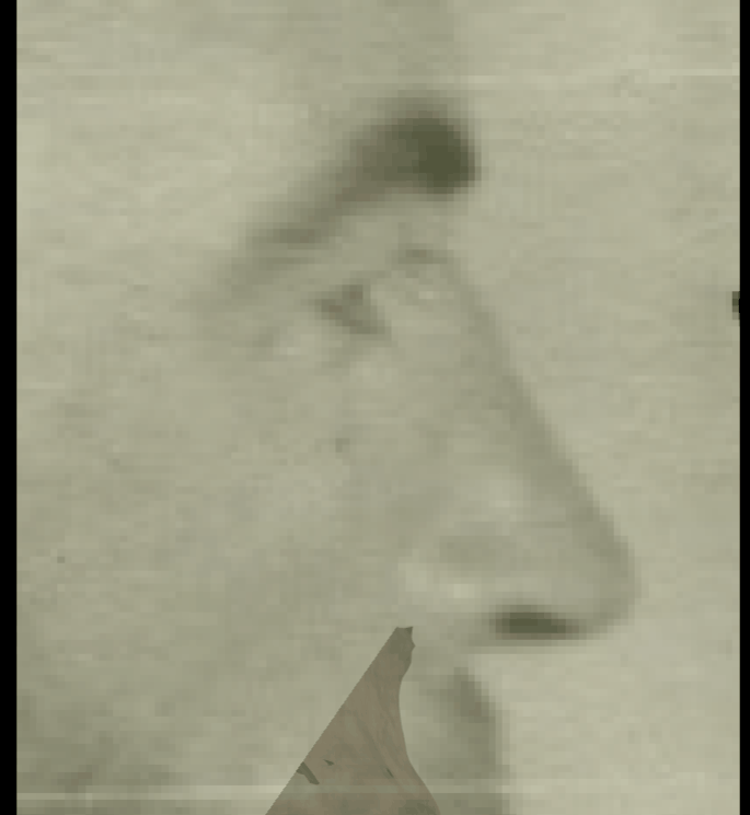 Figure 39. Example of a positive match in which the outline of the nasal bone is evaluated with the transparency and wipe tools, showing that the outline of the nasal bone follows the outline of the nose in the skull with minimal tissue thickness allowance in a consistent way. The wipe tool has been used to show a gradient from the nasal spine to nasion showing consistency in the outlines of both anatomical structures.
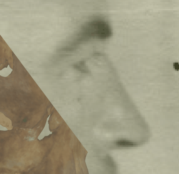 Figure 40. Example of a negative match in which the outline of the nasal bone is evaluated with the transparency and wipe tools, showing that the outline of the nasal bone does not follows the outline of the nose in the skull with minimal tissue thickness allowance in a consistent way. The wipe tool has been used to show a gradient from the nasal spine to nasion showing inconsistency in the outlines of both anatomical structures. In this case the nasal bone morphology is different from the nose in the face and protrudes over the nose contour.
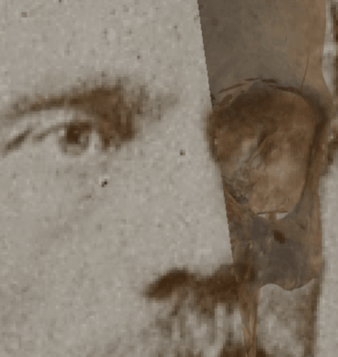 Figure 41. Example of a positive match in which the outline of the nasal bone is evaluated with the transparency and wipe tools, showing that the outline of the nasal bone follows the outline of the nose in the skull with minimal tissue thickness allowance in a consistent way. The wipe tool has been used to show a gradient from the nasal spine to nasion showing consistency in the outlines of both anatomical structures.
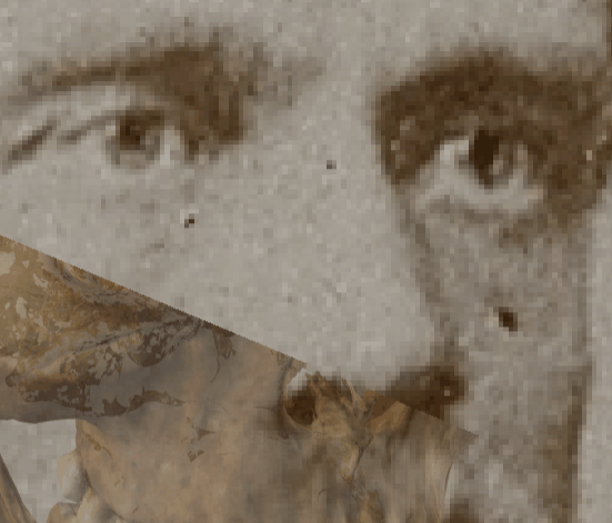 Figure 42. Example of a negative match in which the outline of the nasal bone is evaluated with the transparency and wipe tools, showing that the outline of the nasal bone does not follows the outline of the nose in the skull with minimal tissue thickness allowance in a consistent way. The wipe tool has been used to show a gradient from the nasal spine to nasion showing inconsistency in the outlines of both anatomical structures. In this case the nasal bone morphology is different from the nose in the face and protrudes over the nose contour.